
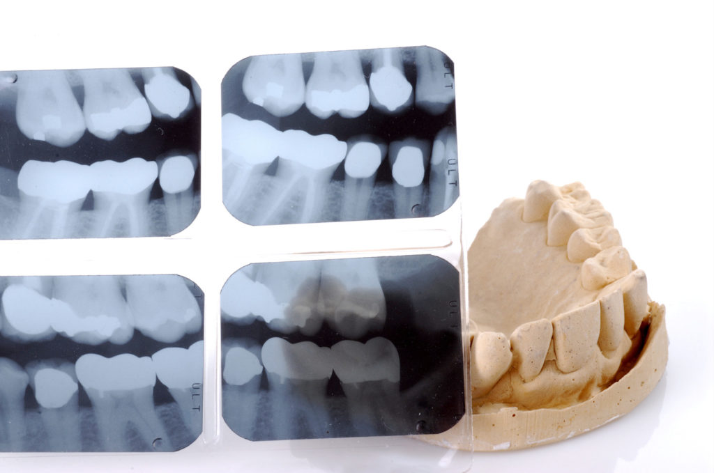
Dental x-rays are a common and essential part of any dental plan. Although the technology associated with dental medicine continues to advance at a rapid pace, a comprehensive set of dental x-rays, or radiographs, remains invaluable when it comes to diagnosing dental issues and creating a comprehensive treatment plan. Coppe + Sears use dental x-rays as a way of seeing the whole picture when it comes to keeping one’s teeth healthy and beautiful. As a diagnostic tool, x-rays allow our doctors to ascertain your oral health by viewing parts of the mouth that are otherwise invisible to the naked eye. Findings can range from simple cavities and tooth decay, to more complex problems such as infections, cysts, and disease.
Despite the importance of x-rays in dentistry, many patients are unsure of their purpose, and understandably want to be certain of their safety. To help you understand more about the role x-rays play in customized dental treatment, we explain below what x-rays are and how we incorporate them into our practice.
The basics of dental x-ray technology
X-rays, also known as radiographs, have a shorter wavelength than visible light and are able to pass through soft tissue in the human body, providing a detailed image of what is going on inside the teeth, bones, and soft tissues of the mouth. The images may diagnose existing problems such as cavities, hidden teeth, or bone loss, as well as indicate the potential for a problem, in order for preventative action to be taken. Intraoral x-rays are the most common type used in dentistry. These show a high level of detail in the tooth, bone, and supporting tissues of the mouth. Dental x-rays may also be used after certain dental treatments for follow-up, to ensure that treatment was successful.
Digital x-rays
Digital dental radiographs are fast becoming an indispensable tool to help dentists and orthodontists better detect, diagnose, treat, and monitor a multitude of oral conditions and diseases. X-ray imaging now predominantly uses digital sensors to replace traditional photographic x-ray film, providing enhanced computer images of the teeth, gums, and other oral structures.
Digital dental x-rays can be taken intraorally or extraorally. Intraoral x-rays are the most common dental x-ray. They provide great detail and are used to detect cavities, check the status of developing teeth, and monitor teeth and bone health. Although extraoral x-rays do not provide the level of detail intraoral x-rays do, and are typically not used to identify individual problems with teeth, they can detect impacted teeth, monitor jaw growth and development, and identify any potential problems between the teeth, jaws, temporomandibular joints, and other facial bones.
Types of intraoral x-rays include the following:
Bitewing x-rays, which show the upper and lower posterior teeth in a single view. This type of x-ray is used to check for any signs of decay between the teeth, as well as how well the upper and lower teeth line up. Bitewing x-rays also help visualize bone loss, should any severe gum disease or dental infection be present.
Periapical x-rays allow the dental professional to look at the entire tooth, from the crown to the the root and on down into the bones that support the tooth. With these x-rays, a dentist can identify dental problems below the gum line or in the jaw, such as impacted teeth, abscesses, cysts, tumors, or the kind of bone changes that are linked to some diseases.
Occlusal x-rays are used to look at the roof or floor of the mouth, and to find extra teeth, teeth that have not yet erupted, jaw fractures, cleft palate, cysts, abscesses, or growths.
Types of extraoral x-rays include the following:
Panoramic x-rays, show the entire mouth, including all of the teeth in the upper and lower arch, in one image. These are used to plan treatment for dental implants, detect impacted teeth (such as canines or wisdom teeth) and jaw problems, and to diagnose bony tumors and cysts. Panoramic radiographs are also used routinely in orthodontics.
Cephalometric projections show the entire head and help dental professionals examine teeth in relation to a patient’s jaw and profile. Orthodontists like Dr. Chad Sears use cephalometric projections to help develop an orthodontic treatment plan for a patient.
Cone beam computerized tomography, or CBCT, shows the body’s interior structures three-dimensionally. CBCT is used to identify problems with the facial bone, such as tumors or fractures. CT scans also are used to evaluate bone for dental implant placement and difficult tooth extractions to avoid possible complications during and after surgical procedures.
Some of the benefits specific to digital dental x-rays are:
- The digital image is processed and available to be viewed immediately, whereas traditional film takes time to be developed.
- There is less radiation needed to produce the same quality image as film. In fact, digital x-rays have up to 80% less exposure to radiation than conventional x-rays.
- Enhancing a digital image is a simple process.
- Digital archiving is more easily attainable.
- Digital radiography is more environmentally friendly.
- Digital images are also easily stored on a computer.
- Digital images can be enlarged and enhanced for review.
- Digital images can be sent electronically for referrals.
Concerns about radiation
We are all exposed to radiation on a daily basis through simple things like smoke detectors, brick homes, and airplane travel, for example. Although too much radiation can be a concern, the additional risk is extremely low in most dental offices, particularly because digital x-rays like those we use at Coppe + Sears can have up to 80% lower doses of radiation than the more traditional films. The amount of radiation exposure from one set of bitewing x-rays is comparable to that of a short domestic flight or the rough equivalent of one day of natural background radiation.
Because digital dental x-rays can provide evidence of decay and other problems early, the benefits of having them done should be weighed against such minimal radiation exposure.
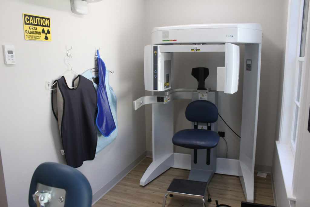
The dental x-ray process
In order to prepare a patient for x-ray exposure, the patient is covered with a heavy lead apron to protect the body from even small amounts of radiation. Next, a small plastic apparatus is inserted into the patient’s mouth and the patient is asked to bite down on it. A member of our team will then take an x-ray picture of the targeted area and will repeat the process until all the necessary images have been obtained. For more information on this process, click here to watch our patient education video on x-rays.
How often are digital dental x-rays necessary for the average patient?
In accordance with the American Academy of Pediatric Dentistry, our doctors will normally take the first comprehensive set of radiographs, which consists of just a few images, between the ages of 3-5. Depending on each patient’s specific case, and additional set of bitewing x-rays is taken every 1-2 years, as well as full mouth scans around ages 8-9 and 17-19.
In general, for those who have no tooth decay and are not at high risk of cavities:
- Adults should have bitewing x-rays done every 2 to 3 years.
- Teens should have bitewing x-rays done every 1½ to 3 years.
- Children should have bitewing x-rays done every 1 to 2 years.
For people who do have tooth decay or are at a high risk of developing cavities:
- Adults should have bitewing x-rays done every 6 months to 1½ years.
- Children and teens should have bitewing x-rays done every 6 to 12 months.
Get the best picture of dental health with the Coppe + Sears Team
Examining one’s mouth for plaque, tartar, and gum disease is a great start toward total dental health, but digital x-rays are the key to diagnosing any unseen problems and designing the best course of treatment to give one healthy teeth and a happy mouth. Our experienced and talented team provides a superior dental and orthodontic experience for families in Lexington and the surrounding areas. To see what digital dental x-rays can tell us about how picture-perfect your smile is, get in touch with us today!